
Tetralogy of Fallot (ToF) is basically a congenital heart defect that affects an estimated 3 in every 10,000 live births. It is a complex heart condition that comprises four distinct structural abnormalities in the newborn’s heart: a ventricular septal defect (VSD), pulmonary stenosis (PS), right ventricular hypertrophy (RVH), and overriding aorta (OA). To help healthcare professionals, medical students, and nurses remember these four components efficiently, we are sharing with you a high-yield mnemonic as the “Tetralogy of Fallot mnemonic” or “ToF mnemonic”.
In this article, we will explore this PROVe mnemonic for Tetralogy of Fallot and its assess its usefulness for doctors. We hope that you find this blog post useful. 🙂
Why is it called Tetralogy of Fallot?
First things first, Tetralogy of Fallot (ToF) is named after the great French physician Étienne-Louis Arthur Fallot, who first described this birth defect in 1888. Fallot observed a peculiar combination of 4 defects in the heart in a series of autopsies he conducted on infants who had tragically died from this cyanotic heart disease (Right to Left shunt). 🙁
Fallot found consistency in all of the autopsies that he conducted. Hence, he named the combination of these four abnormalities as “Tetralogy of Fallot.” The name has since been widely adopted in medicine and is still used today to describe this congenital defect of the heart.
4 Defects of Tetralogy of Fallot
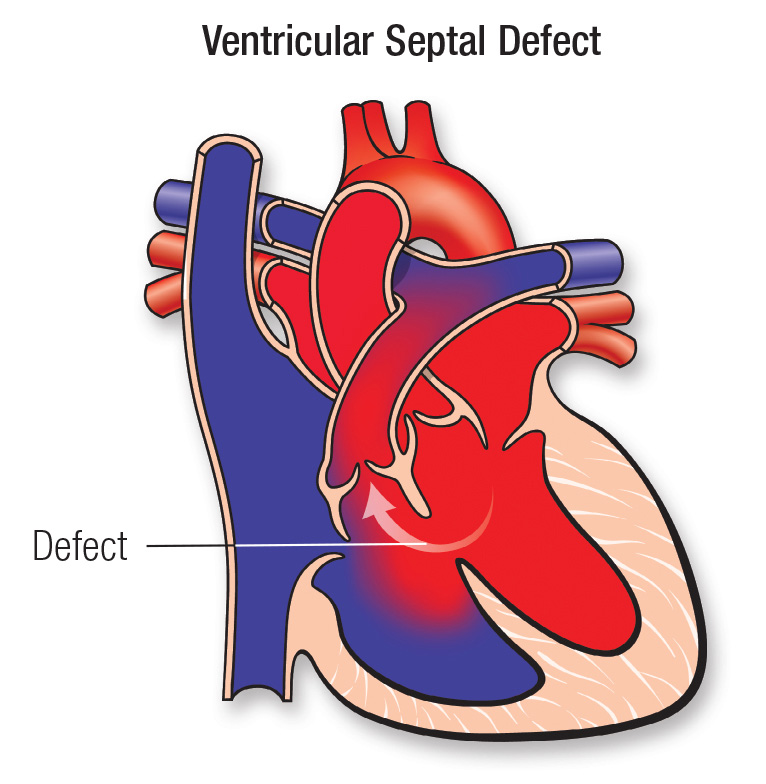
1. Ventricular septal defect (VSD)
|
A hole in the wall of the heart that separates the right and left ventricles. |
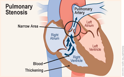
2. Pulmonary stenosis (PS)
|
A condition where the pulmonary valve or artery becomes narrowed, making it difficult for blood to flow from the heart to the lungs. |
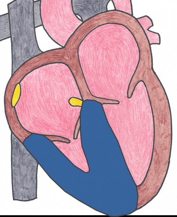
3. Right ventricular hypertrophy (RVH)
|
An enlargement or thickening of the right ventricle of the heart. |
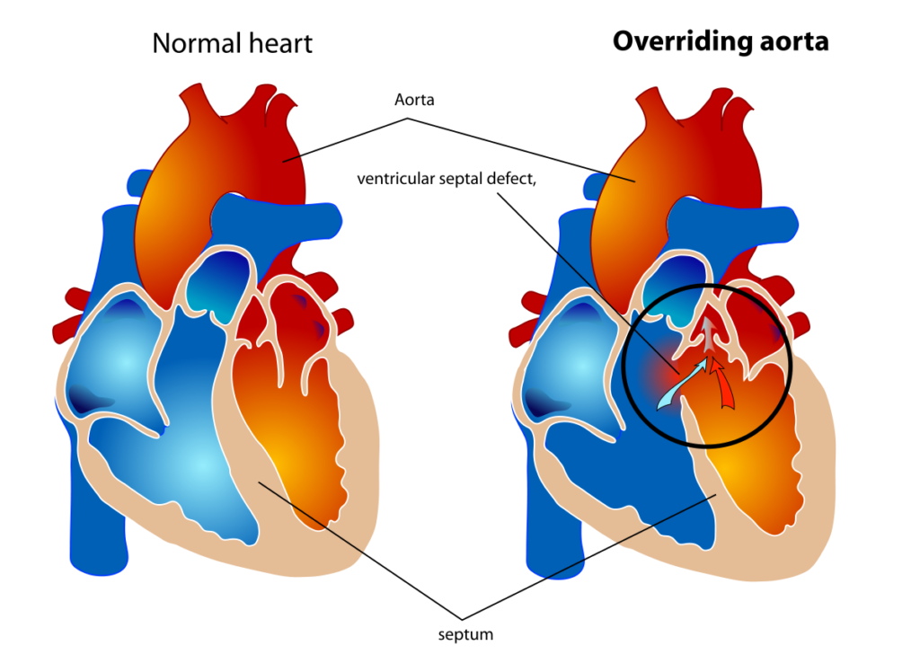
4. Overriding aorta (OA)
|
A condition where the aorta is positioned over both ventricles, rather than just the left ventricle, which results in the mixing of oxygenated and deoxygenated blood which causes cyanosis (bluish discoloration). |
Tetralogy of Fallot Symptoms
The symptoms of Tetralogy of Fallot can include:
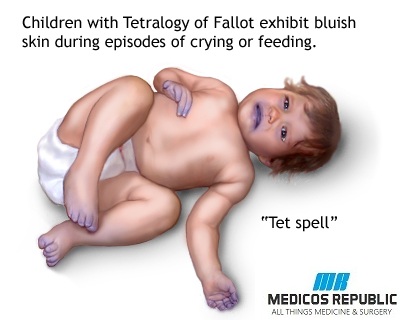
- Cyanosis – defined as a bluish tint to the skin, lips, and nails because of decreased oxygen levels in the blood
- Shortness of breath (SOB)
- Fainting or dizziness
- Clubbing of fingers and toes – a rounding and thickening of the fingertips and toes due to decreased oxygen levels to the tissues of the nail bed.
- Poor weight gain or growth retardation in ToF
Tetralogy of Fallot Mnemonic
1. Tetralogy of Fallot PROVe Mnemonic
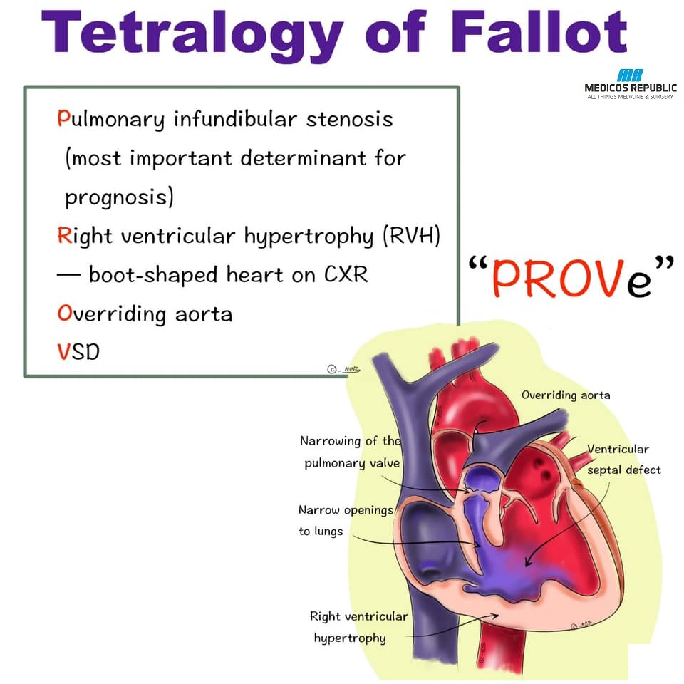
- P – Pulmonary Stenosis
- R – Right ventricular hypertrophy
- O – Overriding of the aorta
- V – Ventricular Septal Defect
[adinserter block=”2″]
2. Tetralogy of Fallot DROP Mnemonic
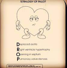
- D – Displaced Aorta
- R – Right Ventricle Hypertrophy
- O – Opening in Septum
- P – Pulmonary valve stenosis
Tetralogy of Fallot CXR Findings
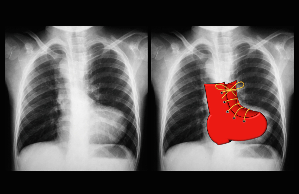
Tetralogy of Fallot chest x-ray findings depend upon the severity of the condition/defect and the age of the patient. However, below are some of the classical CXR findings that may be seen in this disease, including:
- Boot-shaped heart – as seen in the chest X-ray above, it occurs due to the hypertrophy of the right ventricular, the heart appears boot-shaped on the x-ray.
- Narrowed pulmonary artery – in some patients, the pulmonary artery becomes narrowed due to pulmonary stenosis. This can cause the pulmonary artery to appear smaller than normal on the chest x-ray.
- Increased pulmonary markings – due to the decreased blood flow to the pulmonary parenchyma (tissue), there may be increased pulmonary markings on the chest x-ray. This is depicted as a “dirty” lung field.
- Right aortic arch – in some cases of Tetralogy of Fallot, the aorta may be positioned to the right of the windpipe (trachea) instead of to the left which is the normal positioning. This is known as a right aortic arch and may also be visible on the radiographic film.
That’s all for today. We hope that you enjoy this article. Happy learning! 🙂


 Management Mnemonic")


")
")


")
")
