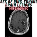
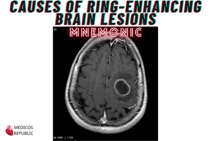
Few things in radiology make medics sit up straighter than a ring-enhancing lesion on brain CT/MRI. It’s the imaging equivalent of your boss walking in mid-ward round — attention-grabbing and usually bad news. 😅
At Sheikh Khalifa Bin Zayed Hospital, Quetta, I’ve seen quite a few such scans — usually followed by intense huddles with Dr. Behroz Rahim (who insists every lesion is just existential angst) or Dr. Bilal Chaudhary (who wants to rule out TB in everyone under 15).
To simplify the differentials, I use the classic — and oddly theatrical — mnemonic:
🎩 “MAGIC DR”
“MAGIC DR” – Causes of Ring-Enhancing Brain Lesions Mnemonic
| Letter | Diagnosis | Key Clinical Clues |
|---|---|---|
| M | Metastases | Multiple lesions, grey-white junction, patient with known or occult malignancy (Lung, Breast, Melanoma). Check with Dr. Danish Ramzan if liver mets are also dancing in the background. |
| A | Abscess | Central restricted diffusion on DWI. Think immunocompromised, poor dental hygiene, or someone who just returned from Mashkel with a fever and altered sensorium. Classic. 😷 |
| G | Glioblastoma | Irregular, solitary lesion with central necrosis. Rapid progression and mass effect. A true villain — and almost always leaves you Googling survival stats. |
| I | Infarct (subacute) | Subacute strokes can enhance in a ring due to breakdown of BBB. Clue: vascular territory distribution and diffusion restriction. Seen this with elderly from Kharan presenting late. 🧓🧠 |
| C | Contusion | Post-trauma, especially in frontal/temporal lobes. Think road traffic accident or a patient referred by Dr. Faisal Afridi after a suspicious “fall from standing height” that needed ortho and neuro both. 🛵 |
| D | Demyelination | Tumefactive MS or ADEM. Ring is usually incomplete (“open ring”). Young adult with visual changes, weakness, and a CT that makes you whisper “please not GBM.” 🧑⚕️🙏 |
| R | Radiation necrosis | History of brain radiotherapy — enhancement due to tissue breakdown. Looks like tumor recurrence but behaves more like your salary after taxes: necrotic and disappointing. 💸 |
🏥 Case from My Ward
A 40-year-old school teacher from Washuk presented with seizures. CT showed a solitary ring-enhancing lesion in the right parietal lobe.
Initial panic: GBM?
MRI: Open ring enhancement.
CSF: Normal.
Final diagnosis (thanks to Dr. Basit Khan’s careful probing): Demyelination — classic tumefactive MS.
Lesson: Not every ring is a curse. Some are just confused inflammation with good PR. 😄
🔬 Clinical Pearl (Exam Gold)
- Diffusion-weighted imaging (DWI) can help differentiate abscess (restricted diffusion) from tumors (typically no restriction).
- Open ring sign → think demyelination.
- If multiple lesions at grey-white junctions → rule out metastases.
- Always ask: Is there trauma, cancer, immunosuppression, or recent radiation? (The four horsemen of ring enhancement.)
Happy learning, folks! 🙂




![Gerstmann Syndrome Features Mnemonic [Easy-to-remember] Gerstmann Syndrome Features Mnemonic](https://www.medicosrepublic.com/wp-content/uploads/2025/06/Gerstmann-Syndrome-Features-Mnemonic-150x150.jpg)
![Cerebellar Signs Mnemonic [Easy to remember] Cerebellar Signs Mnemonic](https://www.medicosrepublic.com/wp-content/uploads/2025/06/Cerebellar-Signs-Mnemonic-150x150.jpg)
![Seizure Features Mnemonic [Easy-to-remember] Seizure Features Mnemonic](https://www.medicosrepublic.com/wp-content/uploads/2025/06/Seizure-Features-Mnemonic-1-150x150.jpg)

![Recognizing end-of-life Mnemonic [Easy to remember]](https://www.medicosrepublic.com/wp-content/uploads/2025/06/Recognizing-end-of-life-Mnemonic-150x150.jpg)

![Multi-System Atrophy Mnemonic [Easy-to-remember] Multi-System Atrophy Mnemonic](https://www.medicosrepublic.com/wp-content/uploads/2025/06/Multi-System-Atrophy-Mnemonic-150x150.jpg)

![How to Remember Southern, Northern, and Western Blot Tests [Mnemonic] How to Remember Southern, Northern, and Western Blot Tests](https://www.medicosrepublic.com/wp-content/uploads/2025/06/How-to-Remember-Southern-Northern-and-Western-Blot-Tests-150x150.jpg)

