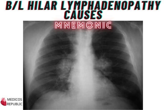
We’ve all seen it — a chest X-ray with those bulky hilar shadows staring back like twin boss fights on either side of the mediastinum. 😮💨
The radiologist writes “bilateral hilar lymphadenopathy” and suddenly, half the differentials in your brain try to exit via the foramen magnum.
But fear not! Like many things in medicine, this too can be tamed — with a trusty mnemonic: CHIASM.
Whether it’s a classic case of sarcoidosis or TB pretending to be mysterious again (as it often does in Balochistan), this memory tool has your back — and both your hila.
📚 Mnemonic: C.H.I.A.S.M — Causes of Bilateral Hilar Lymphadenopathy
| Letter | Cause | Key Points |
|---|---|---|
| C | Carcinoma (esp. lymphoma) | Lymphoma is the #1 neoplastic culprit. Think B symptoms, mediastinal widening, and a refusal to play by textbook rules. Had a classic case last week from Mashkel. CT looked like a Christmas tree. 🎄 |
| H | Histoplasmosis | Fungal drama queen. Rare but loves to mimic TB and sarcoid. A positive travel history or spelunking in chicken coops is your diagnostic compass. 🐓 |
| I | Infection (TB) | Always a suspect in Quetta. If it’s not TB, it’s TB in disguise. Especially common if the patient is from Zhob and brought their own chest X-ray from a local “X-Ray Clinic & Chicken Feed Store.” 🏥🐔 |
| A | Anthracosis | Coal dust exposure — basically lungs saying, “I’ve had enough of this job.” Common in people who worked in brick kilns or dusty construction in Hub or Mach. CXR shows black hilar hilarity. 🪨 |
| S | Sarcoidosis | The king of symmetric hilar lymphadenopathy. Look for the “potato node” hilar enlargement and the classic Löfgren’s triad (hilar LAD + erythema nodosum + arthritis). Dr. Behroz Rahim once diagnosed it before the labs came in — and hasn’t let anyone forget it. 🧠 |
| M | Metastasis | Rarely bilateral, but don’t count it out — testicular, breast, thyroid, and renal primaries can go rogue. Especially in those mysterious “weight loss but fine otherwise” patients. 🧬 |
🏥 Real Case from Quetta
One of my most memorable cases came from Dera Bugti — a 40-something schoolteacher with a dry cough and vague fatigue. X-ray? Bilateral hilar shadows.
Initial bet among the team: TB.
Dr. Basit Khan: “It’s TB till proven otherwise.”
Dr. Faisal Afridi: “Could it be lymphoma?”
Dr. Behroz Rahim: “Check for depression too, just in case.”
Turns out, ACE levels were elevated. Biopsy confirmed sarcoidosis. Moral of the story? Don’t trust TB blindly — sometimes the hilar shadows are just… hilariously misleading.
Happy learning, folks! 🙂
Authored by:
Dr. Aurangzaib Qambrani
MBBS, PLAB, MRCP-UK
Sheikh Khalifa Bin Zayed Hospital, Quetta (Balochistan)






![Gerstmann Syndrome Features Mnemonic [Easy-to-remember] Gerstmann Syndrome Features Mnemonic](https://www.medicosrepublic.com/wp-content/uploads/2025/06/Gerstmann-Syndrome-Features-Mnemonic-150x150.jpg)
![Cerebellar Signs Mnemonic [Easy to remember] Cerebellar Signs Mnemonic](https://www.medicosrepublic.com/wp-content/uploads/2025/06/Cerebellar-Signs-Mnemonic-150x150.jpg)
![Seizure Features Mnemonic [Easy-to-remember] Seizure Features Mnemonic](https://www.medicosrepublic.com/wp-content/uploads/2025/06/Seizure-Features-Mnemonic-1-150x150.jpg)

![Recognizing end-of-life Mnemonic [Easy to remember]](https://www.medicosrepublic.com/wp-content/uploads/2025/06/Recognizing-end-of-life-Mnemonic-150x150.jpg)

![Multi-System Atrophy Mnemonic [Easy-to-remember] Multi-System Atrophy Mnemonic](https://www.medicosrepublic.com/wp-content/uploads/2025/06/Multi-System-Atrophy-Mnemonic-150x150.jpg)

![How to Remember Southern, Northern, and Western Blot Tests [Mnemonic] How to Remember Southern, Northern, and Western Blot Tests](https://www.medicosrepublic.com/wp-content/uploads/2025/06/How-to-Remember-Southern-Northern-and-Western-Blot-Tests-150x150.jpg)

