Pulmonary nodules — the “small round thing” that shows up on CT and sends half the ward into a frenzy.
Is it TB? Cancer? A rogue fungus? Or just a random granuloma left behind by a past infection that didn’t get the memo?
In the middle of this diagnostic chaos, we need a calm, structured approach. That’s where our favorite exam-oriented mnemonic comes in handy: “GAMES”.
Because let’s be honest — figuring out a solitary pulmonary nodule sometimes feels like playing a game of Minesweeper. 💣
🧠 Mnemonic: “GAMES” — Causes of Pulmonary Nodules
| Letter | Cause | Key Notes |
|---|---|---|
| G | Granuloma (TB, fungal) | By far the most common cause in Balochistan. Think TB until proven otherwise. Especially when the patient is from Awaran or Zhob and has weight loss + cough + a story that starts with “6 months ago…” 😷 |
| A | AV Malformation | Rare but dramatic. Hemoptysis + clubbing. Pulmonary angiogram is the MVP here. Dr. Imran Baloch once picked this on echo — legend. 🔍 |
| M | Metastasis | Multiple nodules? Think renal, breast, colon, or choriocarcinoma. Don’t let age fool you — I’ve seen a 35-year-old from Dalbandin present with cannonball mets. 🎱 |
| E | Epithelial Tumor (e.g., carcinoid) | Classic for central nodules. Slow-growing, may present with hemoptysis or a bronchial obstruction. Bonus if the CT shows popcorn calcification. 🍿 |
| S | Sequestration | A congenital beast. Non-functioning lung segment with systemic arterial supply. Not your usual suspect, but Dr. Danish Ramzan loves these. Always CT angio to confirm. 🧬 |
📍 Clinical Experience from Quetta
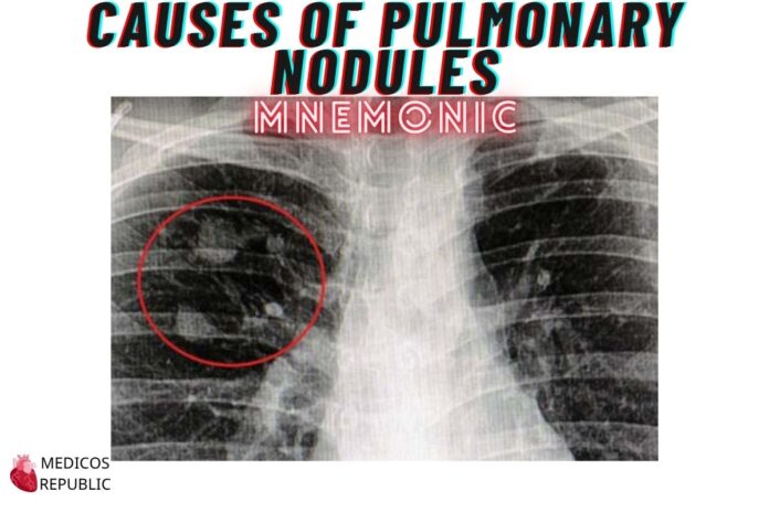
Last month, I had a middle-aged schoolteacher from Washuk who came in with persistent dry cough and fatigue. Chest X-ray showed a solitary RUL nodule.
Dr. Basit Khan suspected TB (as always) and started workup — but the sputum was negative. CT showed a well-defined lesion with central necrosis. Biopsy? Fungal granuloma — Histoplasma, of all things. 😳
Lesson? G in GAMES is still the GOAT in Balochistan.
That’s all for today… happy learning, my friends! 🙂






![Gerstmann Syndrome Features Mnemonic [Easy-to-remember] Gerstmann Syndrome Features Mnemonic](https://www.medicosrepublic.com/wp-content/uploads/2025/06/Gerstmann-Syndrome-Features-Mnemonic-150x150.jpg)
![Cerebellar Signs Mnemonic [Easy to remember] Cerebellar Signs Mnemonic](https://www.medicosrepublic.com/wp-content/uploads/2025/06/Cerebellar-Signs-Mnemonic-150x150.jpg)
![Seizure Features Mnemonic [Easy-to-remember] Seizure Features Mnemonic](https://www.medicosrepublic.com/wp-content/uploads/2025/06/Seizure-Features-Mnemonic-1-150x150.jpg)

![Recognizing end-of-life Mnemonic [Easy to remember]](https://www.medicosrepublic.com/wp-content/uploads/2025/06/Recognizing-end-of-life-Mnemonic-150x150.jpg)
![Multi-System Atrophy Mnemonic [Easy-to-remember] Multi-System Atrophy Mnemonic](https://www.medicosrepublic.com/wp-content/uploads/2025/06/Multi-System-Atrophy-Mnemonic-150x150.jpg)

![How to Remember Southern, Northern, and Western Blot Tests [Mnemonic] How to Remember Southern, Northern, and Western Blot Tests](https://www.medicosrepublic.com/wp-content/uploads/2025/06/How-to-Remember-Southern-Northern-and-Western-Blot-Tests-150x150.jpg)

