Histology is a minor subject of Anatomy during 1st and 2nd Year MBBS. It’s often feared by students because of being graphical and intricate arrangement of subtle biological structures — making it hard to memorize. In order to score well in spotting of histology slides, one must practice reviewing the slides over and over again and memorize the identification points as well.
**PRO TIP: Use histology flash cards or download below-mentioned histology slides in PDF on your phone and practice reviewing before the test. This is one tested and proven method for mastering histology spotting. Hope this works for you as well! 🙂
PAGE JUMPS:
- 1st Year MBBS Histology Slides With Identification Points
- 2nd Year MBBS Histology Slides With Identification Points
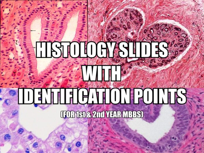
In this blog post, we’re going to share histology slides with identification points for both 1st and 2nd year MBBS classes. The histology slides can be found below in a downloadable PDF file which you can use on your phone or tablet for quick reviewing and test preparation.
Happy learning! 🙂
1st Year MBBS Histology Slides With Identification Points
Below are the 28 histology slides / identification points of 1st Year MBBS:
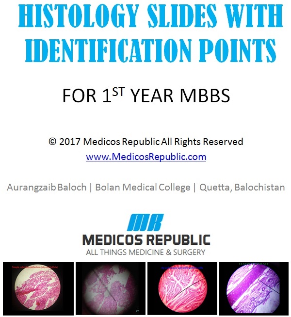
** Click here to view the 1st Year MBBS Histology Slides on Google Drive **
1. SIMPLE SQUAMOUS EPITHELIUM
- Appearance: Flattened cells
- Surface view: rounded central nucleus
2. SIMPLE CUBOIDAL EPITHELIUM
- Cuboidal cells
- Central rounded nuclei
3. STRATIFIED CUBOIDAL EPITHELIUM
- Multilayered Basal cells: high cuboidal or low columnar cells
- Surface layer: cuboidal
4. SIMPLE COLUMNAR EPITHELIUM WITH GOBLET CELLS
- Elongated column shaped cells (height is greater than width)
- Elongated basal nuclei
5. STRATIFIED SQUAMOUS NON KERATINIZED EPITHELIUM
- Surface squamous flattened cells
- No keratin
6. SIMPLE COLUMNAR EPITHELIUM
- Single layer of elongated cells
- Height of cell is greater than width of cell with elongated basal nuclei
7. PSEUDO STRATIFIED COLUMNAR EPITHELIUM
- Single layer of cells
- Nuclei lying at different levels; give the false appearance of “stratified”epithelium
8. FIBRO CARTILAGE
- No perichondrium
- Chondrocytes within lacunae, arranged in rows
9. ELASTIC CARTILAGE
- Perichondrium is present
- Elastic fibres in matrix
- Chondrocytes in lacunae in the form of isogenous groups
10. HYALINE CARTILAGE
- Dense Irregular CT (perichondrium)
- Chondrocytes (in the form of isogenous groups) within lacunae
- Visibility to naked eye: Bluish white translucent appearance
11. THIN SKIN
- Stratified squamous keratinized epithelium
- Hair follicles and roots present
- Sebaceous and sweat glands present
12. THICK SKIN
- Stratified squamous keratinized epithelium
- No hair roots and follicles
- Pacinian corpuscles
14. DENSE REGULAR CONNECTIVE TISSUE
- Parallel collagen fibers in the form of bundles
- Compressed elongated fibroblasts between the collagen fibers
15. MUCOUS ACINI
- Pyramidal cells
- Flattened nuclei towards the base
16. SPINAL CORD
- Central canal
- Butterfly shaped grey matter
- Dorsal median sulcus and ventral median fissure (depending on the portion under examination)
17. ARTICULAR CARTILAGE
- Superficial tangential layer
- Intermediate transitional layer
- Radial zone
- Calcified zone
18. COMPACT BONE
- Haversion Canal
- Lamellae
- Osteocytes in lacunae
19. SPONGY BONE
- Trabeculae
- Red marrow
20. CARDIAC MUSCLE
- Striated appearance
- Intercalated discs
21. SMOOTH MUSCLE
- Non-striated appearance
- Single central nucleus
- Spindle shaped cells
22. SKELETAL MUSCLES
- Characteristic striated appearance
- Peripheral nuclei
- Nucleus ovoid in shape
23. ADIPOSE TISSUE (WHITE)
- Unilocular
- Nuclei towards periphery
24. ELASTIC ARTERY
- No distinct outer and inner elastic membranes
- Tunica adventitia: merges with CT, contains vasa vasorum
- Tunica media: elastic fibres are present in a large amount
25. SPLEEN
- Red pulp: splenic cord and venous sinuses
- White pulp: contains lymphatic nodules and germinal centre
26. THYMUS
- Medulla is lightly stained
- Hassall’s corpuscles in medulla
27. LUNG
- Alveoli: Type I alveolar cells (simple squamous epithelium); Type II alveolar cells (cuboidal cells)
- Respiratory bronchioles: lined by simple cuboidal epithelium
28. TONSIL
- Stratified squamous non keratinized epithelium
- Crypt
- Trabeculae
- Lymphatic nodules
2nd Year MBBS Histology Slides With Identification Points
Below are the 58 histology slides / identification points of 2nd Year MBBS:
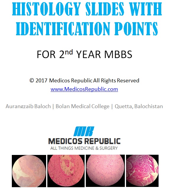
** Click here to view the 2nd Year MBBS Histology Slides on Google Drive **
1. DUCTUS DEFERENS
- Irregular Convoluted single lumen
- Low columnar pseudostratified epithelium with stereocilia
- Very thick (3 layered) muscularis
2. KIDNEY CORTEX
- Simple SQ. Epithelium
- Bowman’s capsule
- Glomerulus
- Proximal and distal convoluted Tubules
3. THIN SKIN
- Epidermis consists of ST. SQ. Keratinized epithelium
- Dermis
- Skin appendages i.e hair follicle, sweat and sabeceaous glands
4. THICK SKIN
- Includes a cartilage i.e hyaline cartilage in appearance
- Above mentioned features of thin skin
- Mention name i.e Vestibule of the nose
5. SPINAL CORD
- Outer white matter
- Inner gray matter which is H shaped
- Centeral canal lined by empendymal cells
6. CEREBELLAR CORTEX (cerebellum)
- Cortex consists of three layers : Outer molecular layer & Inner granular layer
- In between these two layers is a single layer of Purkinjee cells
- Inner white matter
- Outter gray matter
7. CEREBRUM
- Comprises of six layers
- 3 types of cells: a. Pyramidal cells b. Granule cells. c. Triangle cells
8. ARTERY
- Lumen is rounded and regular
- Tunica Intima
- Tunica media => Endothelium :: Simple SQ. Epithelium
- Tunica externa
9. VEIN
- Irregular lumen
- Thin & regular tunica media
10. THYMUS
- Thymic lobules
- Hassel’s corpuscles
- Complete and incomplete septa and lobules
- Trabaculae
11. HYALINE CARTILAGE
- Perichondrium
- Isogenous Chondrocytes in Lacunae
- Hylose
- Cell nest
- Collagen fibers present but not visualized
12. ELASTIC CARRILAGE
- Perochondrium
- Chondrocytes scattered in matrix
- Elastic fibers present and visualized
13. LIP
- Dermal Papillae along the foldings
- Core of skeletal muscles
- Labial Salivary glands
14. PENIS
- Corpus Cavernosum
- Corpus spongiosum
15. SPONGY BONE
- Few Haversion System
- Osteocytes with their lacunae
- Bony trabaculae
- Periosteum
- Endosteum
16. SKELETAL MUSCLE
- Cross striations present
- Multinucleated
- Nuclei at the periphery
17. CARDIAC MUSCLES
- Striated muscle fibers with intercalated discs
- One or two oval central nuclei
18. SMOOTH MUSCLES
- Spindle shaped muscle fibers
- Flattened centrally arranged nucleus.
19. PALANTINE TONSIL
- Partial epithelium & partial capsule
- Tonsillar crypts
- ST. SQ. Nonkaretinized epthelium
- Lymphatic nodules
- Aggregation of lymphocytes
20. LYMPH NODES
- Outer cortex having lymphatic nodules with Germinal centers
- Inner medulla having medullar sinuses
21. SPLEEN
- Bilirth cords and venous sinuses
- White pulp
- Red pulp
- Peri-arterial sheeth
22. THYMUS
- Thymic Lobules
- Hassel’s corpuscles
- Complete and incomplete septa and lobules
23. TOUNGE
- ST. SQ. Nonkeratinized
- Lingual papillae
- Taste buds
- Mucous and serous acini
24. ESOPHAGUS
- All layers: Mucosa, Submucosa, Muscularis Externa and Adventitia
- Mucosa lined by ST. SQ. Nonkeratinized
25. STOMACH
- Irregular Mucosal Folds: Rogue (only specific for stomach)
- Mucosa lined by Simple Columnar Epithelium
- Gastric pits
- Gastric ducts
- Pareital cells and cheif cells
26. DUODENUM
- Mucosa has “Leaf” like out folding
- Lined by Simple Columnar Epithelium
- Brunner’s gland in sub-mucosa
27. JUJENUM
- Mucosa has “finger” like out folding
- Lined by Simple Columnar Epithelium
- Lymphatic nodules
28. ILEUM
- Club-shaped villi
- Lined by Simple Columnar Epithelium
- Payer’s patches
29. LARGE INTESTINE
- All layers: Mucosa, Submucosa, Muscularis Externa and Adventitia
- Mucosa in form of Crypts
- Abundance of Goblet cells
- No villi
- Tenia colai
30. GALL BLADDER
- Simple columnar epithelium
- Wall is composed of: Mucosa, muscularis externa and adventitia
- Rogue (outward projections) and diverticulum (inward fold)
- Sub-mucosa absent
30. SALIVAR GLANDS
- Mixed glands both mucous and serous acini. Predominantly mucous
- Striated, Intercalated and interlobular duct
- Serous demilunes
31. SUB MANDIBULAR GLANDS
- Mixed glands both mucous and serous acini. Predominantly serous
- Striated, interclalated and interlobular ducts
32. PAROTID GLAND
- Pure serous acini are present
- Striated, Intercalated and interlobular ducts
33. LIVER
- Hepatic sinusoids
- Portal canal
- Hepatocytes
34. PANCREASE
- Pancreatic accini
- Pancreatic islets
- Pacinian corpuscles
35. THYROID GLAND
- Thyroid follicles lined by simple cuboidal epithelium
- Follicle with collide
- Simple cubodial epithelium
- Parafollicular cells
36. ADRENAL GLAND
- It is divided into outer cortex and inner medulla
- Adrenal cortex is composed of 3 Zones:
A. Outer: Zona Glomerulosa
B. Middle: Zona Fasciculate
C. Inner: Zona Reticularis
37. TRACHEA
- C-shaped rings of hyaline cartilage
- Pseudostratified ciliated epithelium
38. LUNGS
- Network of Alveoli and alveloar ducts
- Intrapulmonary bronchus lined by Pseudostratified epithelium
- Respiratory bronchioles
- Bronchi with hyaline cartilage
39. KIDNEY
- Cortex has renal corpuscles
- Medulla has papillae made up if collecting ducts
- Glomeruli
- Convoluted tubules
40. URETER
- Star like lumen
- Epithelium: Transitional Epithelium
41. URINARY BLADDER
- Epithelium: Transitional Epithelium
- Smooth muscles bundles are present
42. TESTIS
- Seminiferous tubules lined by St. Cuboidal Epithelium
- Lydig cells
- Germinal epthelium
- Tunica Albogenia
- Tunica Vasculosa
43. VAGINA
- Epithelium: St. Sq. Nonkeratinized epithelium
- Lamina propria lacks glands
- Bundles of smooth muscles
44. UTERUS
- Wall is composed of 3 layers Endometrium, Myometrium and Perimetrium
- Germinal epthelium
45. FILLOPIAN TUBE
- Simple columnar epithelium
- Wall has three layers: Mucosa, Muscularis Externa and Serosa
- Irregular mucosal folds
46. OVARY
- Different stages of the graffian follicles
- Corpus luetum
- Primary follicle
- Cortical stroma
- Germinal epithelium
47. MAMMARY GLANDS
- Lactiferous ducts
- Alevoli with milk
- Acini appears round spherical
48. VAGINA
- Stratified Squamous non-kertanizied
- Mucosal Folds
- Smooth muscles
49. CORNEA
- Corneal stroma
- Desment’s layer
- Corneal epithelium: St. Sq. Non-Keratinized Epithelium
50. RECTUM
- Tenia coli absent
- Simple Columnar Epithelium
- Pyares patches
- Numerous goblet cells
51. PROSTATE
- Capsule
- Prostatic glands
- In the lumen deposition of carbs and protiens: Corpora Amyleca
52. APPENDIX
- Lymphatic nodules
- Goblet cells
- Irregular lumen
53. COLON
- Tenia coli
- Numerous Goblet cells
- Villi absent
54. EPIDIDYMUS
- Pseudostratified columnar epithelium
- Speematozoa in lumen of the duct
55. SEMINAL VESICLE
- Highly convoluted lumen with crypts and cavities
- Pseudostratified epithelium
56. RETINA
- Distinct 10 layers can be recognized
- Rods and cones are present
- Pigment epithelium
57. SUBLINGUAL GLANDS
- No capsule present
- Predominantly Mucous acini with few serious one
- Few serious demilunes are present
- No Intercalated ducts
58. SUBMANDIBULAR GLANDS
- Mixed alveoli
- Many serious demilunes
- Shorter and narrower intercalated ducts

![How to Survive 1st Year MBBS of Medical College? [Complete Guide]](https://www.medicosrepublic.com/wp-content/uploads/2017/08/How-to-Survive-1st-Year-MBBS-of-Medical-College-218x150.jpg)

![Imaging Anatomy: Knee, Ankle, Foot 2nd Edition PDF Free Download [Direct Link]](https://www.medicosrepublic.com/wp-content/uploads/2018/12/Imaging-Anatomy-Knee-Ankle-Foot-2nd-Edition-PDF-Free-Download-150x150.jpg)
![Burns’ Pediatric Primary Care PDF Free Download [Direct Link] Burns' Pediatric Primary Care PDF](https://www.medicosrepublic.com/wp-content/uploads/2024/02/Burns-Pediatric-Primary-Care-PDF-150x150.jpg)
![Color Textbook of Histology PDF Free Download [Direct Link] Color Textbook of Histology PDF](https://www.medicosrepublic.com/wp-content/uploads/2024/03/Color-Textbook-of-Histology-PDF-Free-Download-1-150x150.jpg)
![Clinical Anatomy: Applied Anatomy for Students and Junior Doctors PDF Free Download [Direct Link] Clinical Anatomy Applied Anatomy for Students and Junior Doctors PDF](https://www.medicosrepublic.com/wp-content/uploads/2022/05/Clinical-Anatomy-Applied-Anatomy-for-Students-and-Junior-Doctors-PDF-Free-Download-150x150.png)
![MBLEx Test Prep 2024-2025 PDF Free Download [Direct Link] MBLEx Test Prep 2024-2025 PDF](https://www.medicosrepublic.com/wp-content/uploads/2024/02/MBLEx-Test-Prep-2024-2025-PDF-150x150.jpg)

![All In The Mind PDF Free Download [Direct Link] All In The Mind PDF](https://www.medicosrepublic.com/wp-content/uploads/2023/04/All-In-The-Mind-PDF-150x150.jpg)
![First Aid Q&A for the USMLE Step 1 3rd Edition PDF Free Download [Direct Link] First Aid Q&A for the USMLE Step 1 PDF](https://www.medicosrepublic.com/wp-content/uploads/2018/03/First-Aid-QA-for-the-USMLE-Step-1-PDF-Free-Download-150x150.jpg)
![A Biologist’s Guide to Analysis of DNA Microarray Data PDF Free Download [Direct Link] A Biologist's Guide to Analysis of DNA Microarray Data PDF](https://www.medicosrepublic.com/wp-content/uploads/2024/03/A-Biologists-Guide-to-Analysis-of-DNA-Microarray-Data-PDF-Free-Download-1-150x150.jpg)
![Sacred and Herbal Healing Beers PDF Free Download [Direct Link] Sacred and Herbal Healing Beers PDF](https://www.medicosrepublic.com/wp-content/uploads/2024/04/Sacred-and-Herbal-Healing-Beers-PDF-Free-Download-1-150x150.jpg)

![Coding Notes: Pocket Coach for Medical Coding PDF Free Download [Direct Link] Coding Notes Pocket Coach for Medical Coding PDF](https://www.medicosrepublic.com/wp-content/uploads/2024/04/Coding-Notes-Pocket-Coach-for-Medical-Coding-PDF-Free-Download-1-100x70.jpg)
![NCLEX: Pharmacology for Nurses PDF Free Download [Direct Link] NCLEX Pharmacology for Nurses PDF](https://www.medicosrepublic.com/wp-content/uploads/2024/04/NCLEX-Pharmacology-for-Nurses-PDF-Free-Download-1-100x70.jpg)
![Med Math Simplified 2nd Edition PDF Free Download [Direct Link] Med Math Simplified 2nd Edition PDF](https://www.medicosrepublic.com/wp-content/uploads/2024/04/Med-Math-Simplified-2nd-Edition-PDF-Free-Download-1-100x70.jpg)
![Medical Terminology Made Simple PDF Free Download [Direct Link] Medical Terminology Made Simple PDF](https://www.medicosrepublic.com/wp-content/uploads/2024/04/Medical-Terminology-Made-Simple-PDF-Free-Download-1-100x70.jpg)